horizontal part of frontal bone
PPT - OSTEOLOGY BONES PowerPoint Presentation, free download - ID:2300393 we have 8 Pics about PPT - OSTEOLOGY BONES PowerPoint Presentation, free download - ID:2300393 like Inferior View of Horizontal Orbital Part of Frontal Bone | Neuroanatomy, Inferior View of the Frontal Bone | Neuroanatomy | The Neurosurgical and also PPT - The Skeleton PowerPoint Presentation, free download - ID:675472. Read more:
PPT - OSTEOLOGY BONES PowerPoint Presentation, Free Download - ID:2300393
 www.slideserve.com
osteology ppt bones powerpoint presentation foramen cecum
PPT - LIF101 Anatomy PowerPoint Presentation, Free Download - ID:1342220
 www.slideserve.com
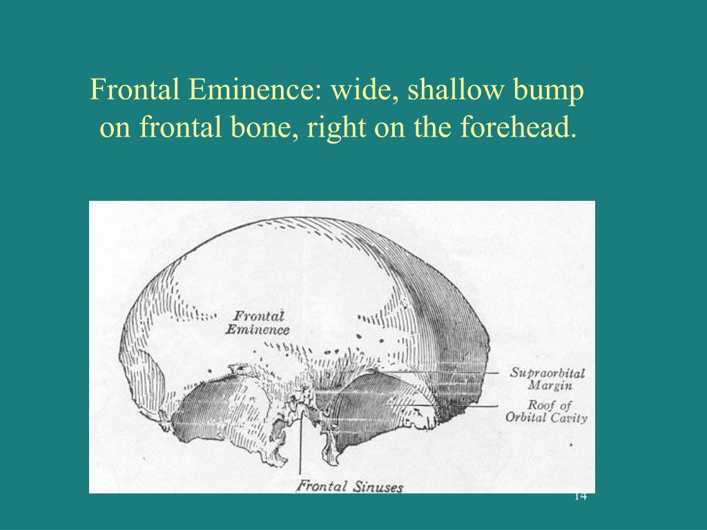
frontal eminence forehead bone bump anatomy shallow wide right ppt powerpoint presentation
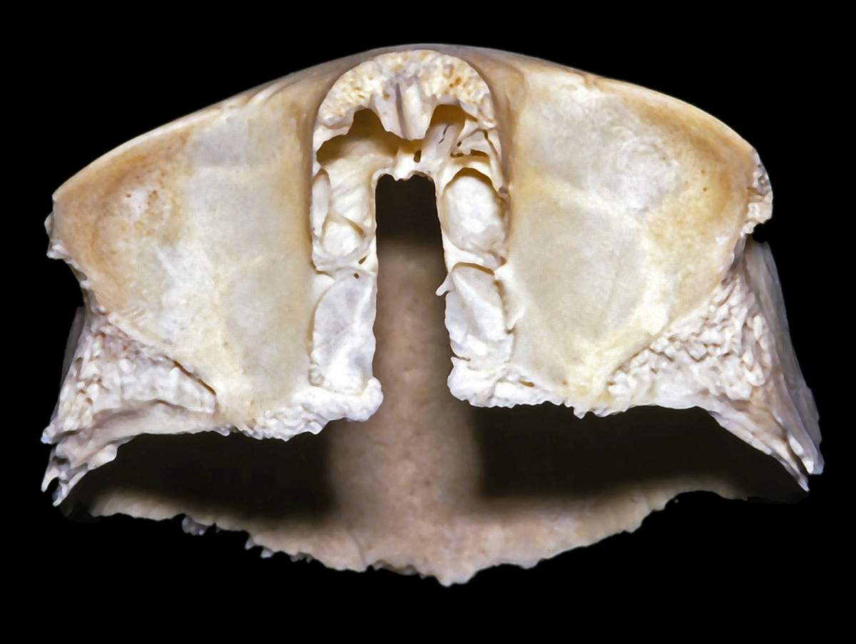
Inferior View Of The Frontal Bone | Neuroanatomy | The Neurosurgical
 www.neurosurgicalatlas.com
neurosurgicalatlas surgical
PPT - The Skeleton PowerPoint Presentation, Free Download - ID:675472
 www.slideserve.com
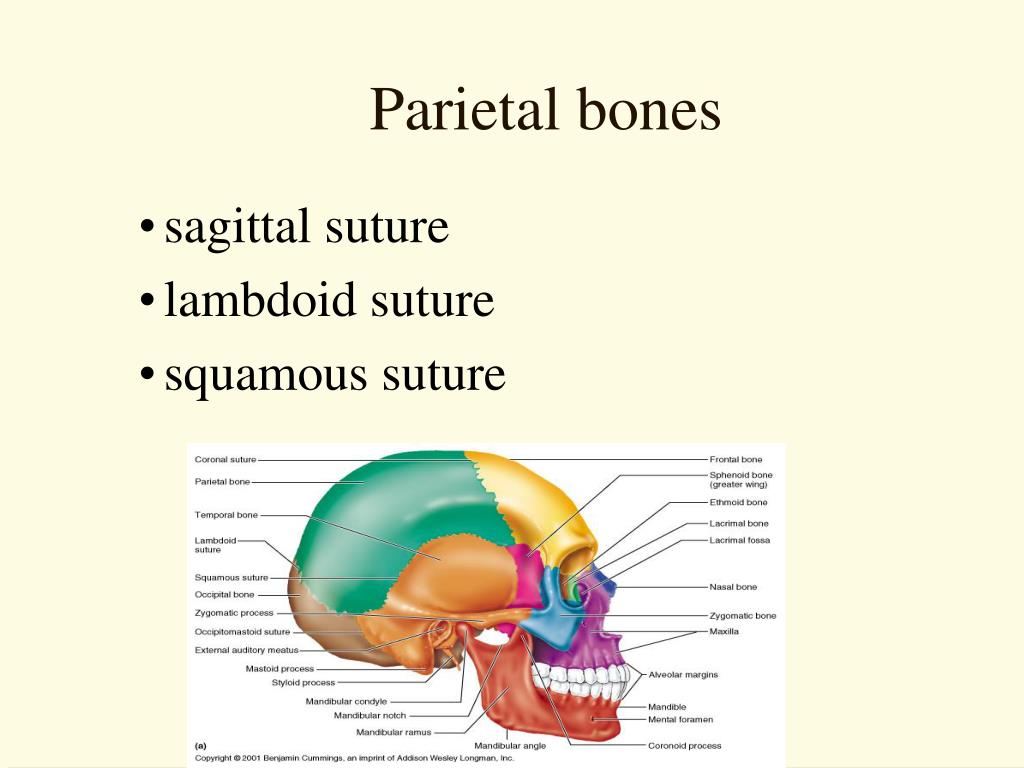
parietal suture squamous lambdoid sagittal
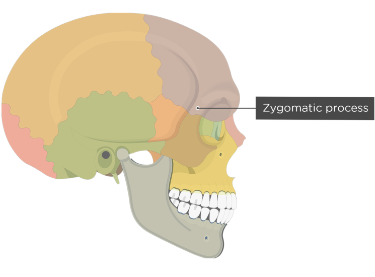
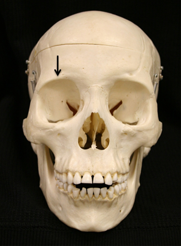
Frontal Bone Anatomy
 www.getbodysmart.com
frontal bone zygomatic process anatomy bones skull cranial
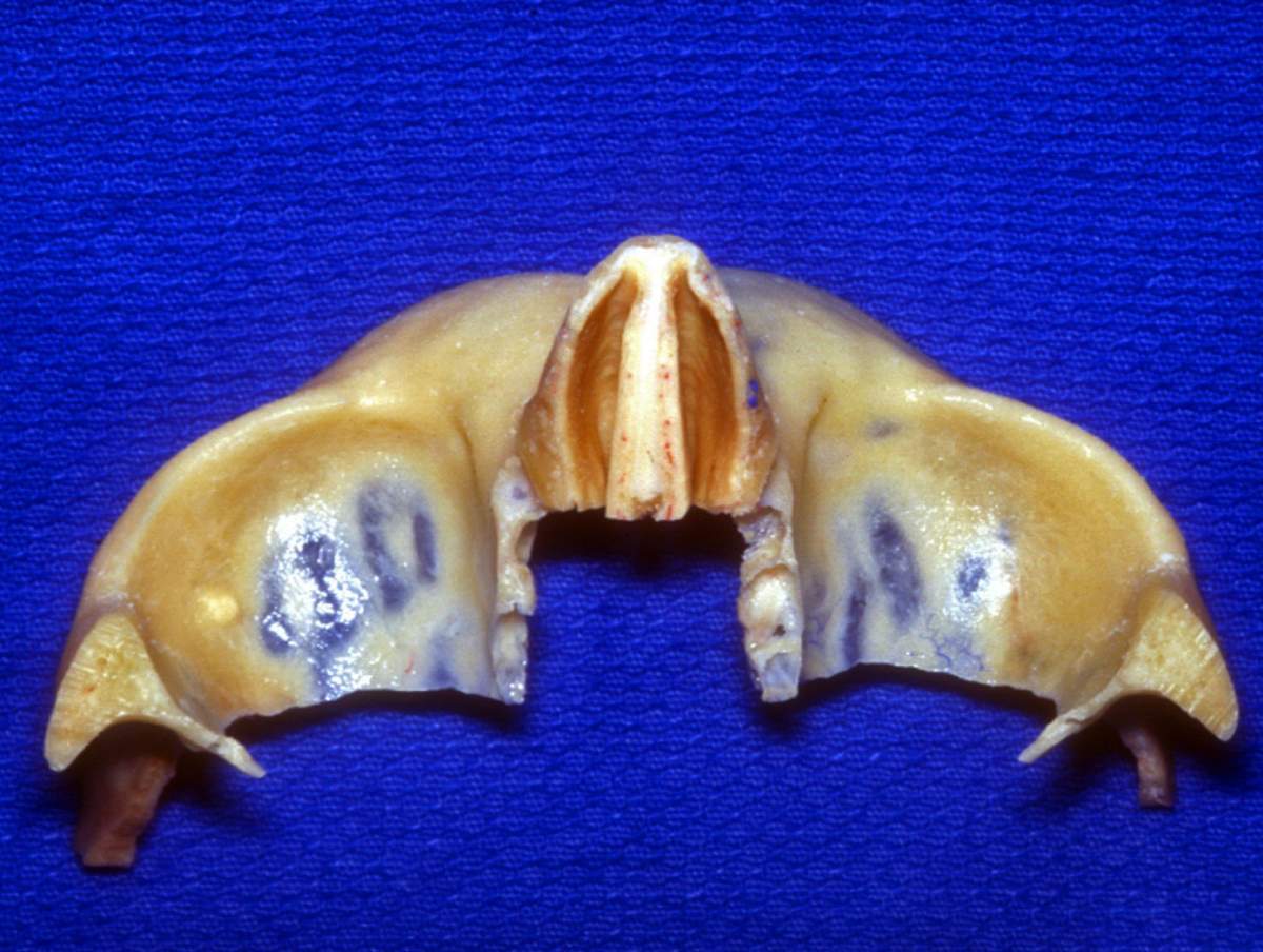
Inferior View Of Horizontal Orbital Part Of Frontal Bone | Neuroanatomy
 www.neurosurgicalatlas.com
bone frontal orbital inferior horizontal
Development Of The Trabecular Structure Within The Ulnar Medial
 anatomypubs.onlinelibrary.wiley.com
coronoid process medial wiley figure ulnar trabecular structure within dogs development young
Lateral Cephalometric Skull Anatomy – Part I – Dr. G's Toothpix
 drgstoothpix.com
skull cephalometric lateral orbital orbit anatomy cavity superior roof
Inferior view of horizontal orbital part of frontal bone. Lateral cephalometric skull anatomy – part i – dr. g's toothpix. Coronoid process medial wiley figure ulnar trabecular structure within dogs development young